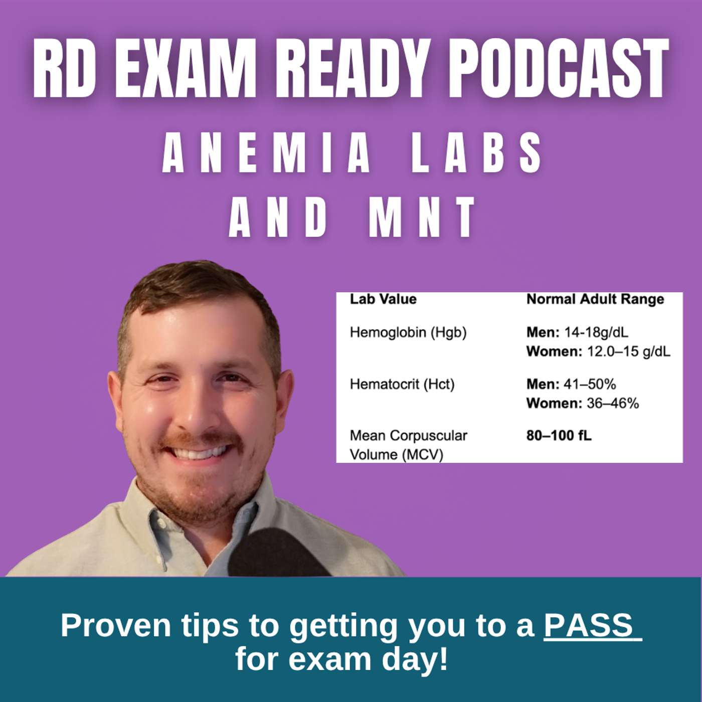
In this podcast episode, host Zach Kaseberg returns from a month-long hiatus caused by a staff strike that forced him to work as a cook for 60-hour weeks. He introduces a discussion on anemias, a key topic for the CDR exam. Anemia is defined as a deficiency in red blood cell size, number, or hemoglobin content. The mean corpuscular volume (MCV) measures red blood cell size: normal is 80-100 fL, microcytic (small) is below 80 fL, and macrocytic (large) is above 100 fL. Microcytic anemia is typically caused by iron deficiency, leading to hypochromic (pale) cells due to low hemoglobin, with symptoms like pale skin, brittle nails, and spoon-shaped nails (koilonychia). Labs show low ferritin and high transferrin. Treatment includes oral iron with vitamin C for absorption, dietary heme iron from meats, and avoiding inhibitors like calcium. Macrocytic anemia results from B12 or folate deficiencies (megaloblastic anemias), causing large, immature cells. B12 deficiency, often due to lack of intrinsic factor, presents with tingling, poor memory, and high homocysteine and methylmalonic acid levels; it is treated with injections or high oral doses. Folate deficiency, common in pregnancy, causes fatigue and glossitis, treated with dietary fruits, vegetables, and fortified grains. A practice question with an MCV of 75 fL confirms iron deficiency, as it is microcytic, ruling out B12 and folate. The episode emphasizes using lab values and dietary patterns to diagnose anemias on the exam.
Hello, RDs and RDs to be. Welcome to the RD exam ready podcast. I'm Zach Kaseberg, your tutor, your coach, your guide along the path to passing the CDR exam. By the biggest passion is to help RDs and RDs to be just like you to note how to effectively pass the CDR exam and if that sounds like you, then hey, you're in the right place. Quick plug here before I get into stuff folks, we're gonna be doing another RD exam ready live on YouTube on May 20th of 2026. So if you're listening live, we're gonna be doing that May 20th, 2026, bringing these back, doing it at seven o'clock, central time. That's gonna be eight o'clock Eastern time, seven o'clock, central time, six o'clock, mountain time, five o'clock Pacific is what that should be there. Yep, perfect. We'll be discussing some common medications for exam day during that session. So if you're listening live, come join us on YouTube on the RD exam ready page or just go with what's in the link here of this podcast description. You gotta see, I've missed teaching you all while I was out on this little short hiatus. Those are you that belong to the mailing list and if you don't, you should 'cause you can get some free content through there. Know that maybe I had one of the weirdest months as an RD. I was taken from my nice cushy desk and I was thrown into our kitchens as a cook and that was due to our culinary staff members going out on strike all of the month of April for 28 days. A very odd month indeed for my life professionally and I'd say on a personal level, it was extremely difficult on my home life. Just having a young infant, having a family, working so many hours per week, doing you know, 60 hours per week, six days a week, very odd shifts and odd times. So it was a whole lot going on and thankfully, that's all over. So for those of you that are listening live, thank you for the patience with me during the short hiatus and if you're listening back years later, then hey, you didn't miss a beat. Since I spent so much time during that time, I worked with a lot of unions during that time, someone a little bit exhausted on the union content, I think we need a little bit of a palate cleanser. So to speak here, let's talk blood today, the real palate cleanser, right folks? When I give some background to you today about different ennemias, the labs associated the deficiencies that we see and typically follow through these. And why I think this is a great topic to discuss is because on the exam, we can generally say that there's gonna be some questions that pop up about ennemias. For instance, it may ask, give you a question that describes a patient's diet, give some associated labs and then ask you to interpret which type of ennemia this patient may be experiencing. Or I could go the other way, maybe it's gonna be a case where they give you the lab and ask you to fit with some type of dietary deficiencies or restriction. For these types of questions, it takes several elements of understanding and it's really gonna take your ability to kind of look at the information present to you and make sure that you understand how you're going to use that for an exam day knowing how you're going to analyze or infer about what type of condition might be associated with those labs and those dietary needs. A lot of these conversations about these labs really have to do with the red blood cell itself, aka the erythrocyte. So let's talk about it today. Ennemia means deficiency in the size or the number of red blood cells or the amount of hemoglobin they contain. Now just a reminder about the red blood cell itself, it contains hemoglobin. This provides the red pigment that is found in red blood cells. Hemoglobin is a conjugate protein, it's carrying four hem groups and a globin molecule. It's the oxygen carrying pigment of the erythrocytes. Iron itself our favorite mineral to talk about is an essential element to hemoglobin. See you can infer that without iron, you are going to lack some of the hemoglobin necessary. I wanna talk about red blood cells even more today. And I wanna talk about the size of them, not the size shame them here, but we gotta talk about that. We're going to discuss the mean corpuscular volume or MCV is I'm gonna call it from here on out. Mean corpuscular volume or MCV describes the size of the red blood cell in this case. On any normal person who doesn't have an enemy of any kind, the mean corpuscular volume or MCV should be between 80 to 100 fento leaders, fento leaders as it's called here. This would be the normal size of a red blood cell. If an MCV is below 80, though it indicate that the cell is micro-cidic, very, very small, very, very small, micro-cidic, break that word down micro small-cidic cell, meaning it's going to be small and ultimately immature. This cell just may not have developed fully to get to the point that it needs to be, and that's why it's going to be a micro-cidic cell. Sometimes this is due to not having enough iron to develop to its normal size. This is why iron typically leads to micro-cidic anemias. The cells are immature and they're small. The other side of that would be an MCV that is above 100. An MCB above 100 would indicate the cell is macro-cidic. Macro-cidic, break that word down macro-big-cidic cell. This is a large red blood cell. This would not be caused by iron, not at all. Rather, this would be caused by a deficiency of vitamin B12 and folic acid. You can think B12 as big B12 or big cell. That can be your little memory trick to know a big cell means B12. I also think of folate with this. I think of folate, I think of these big, big red blood cells, and then I think of a pregnant woman's belly. Large belly due to the baby, not the folate. The baby that's growing inside there. So I think of folate, I think of baby, I think of mom, and I think of a big pregnant belly as a big, a re-through site's cell. So that's my little memory trick for going through that. Now, I want to swing back into hemoglobin just a little bit. Your hemoglobin labs will have different lab values, depending on a fewer male or female. A man should have a hemoglobin that's right between 14 to 18 grams per desoliter, and a woman should have a hemoglobin that's going to be between 12 to 15 grams per desoliter. My best way of remembering this is thinking about when a lot of kids go through puberty. For boys, they're always a little later than girls, right? So generally, we can say 14 to 18 years old is when boys go through puberty. And for girls, typical puberty age might be a little bit earlier than boys. Maybe we see something more like 12 to 15 years old might be what we see for girls. So we just remember those numbers. 14 to 18 for boys, 12 to 15 for girls, and we have our numbers nice and dialed in. Now, if, girl, boy, whoever, let's say your hemoglobin is below that threshold, that would indicate that there's a low amount of hemoglobin in the blood, and that's a problem. Low hemoglobin would likely be due to a low amount of iron, but could be caused by a low amount to be 12 or fully as well. If you have an iron deficiency, you likely have a low hemoglobin because iron is so important for the development of hemoglobin. So if you're missing that very essential core that iron provides, you're going to be missing some of the hemoglobin in your blood. When we see low hemoglobin, we're likely going to be thinking of an iron deficiency, but not necessarily in all cases. We can lose blood and have a low hemoglobin in so many ways. You can lose blood in lots of ways. You could have a gunshot. That's one way you would lose blood from outside your body, something like that. You could be losing blood inside your body. You could have a gastric ulcer, a bleeding ulcer of some kind. You could be going through menstruation, for instance, which could cause you to lose blood in that case. So these are all ways in which hemoglobin can be lost in the body. When we're talking about an erythrocyte, we also need to refer to the color of those erythrocytes. So now this is directly to do with the hemoglobin because hemoglobin contributes the red color in a red blood cell. A normal red blood cell that is rich in red color and red is known as normo-chromic. Normo-chromic is what we call that. Normo-meaning-normal, chromic-meaning-color. So that would be a normal red blood cell in that case. When there is low levels of color or their pale, we call that hypo-chromic-hypo, meaning low, chromic-meaning-color. In an iron deficiency anemia, we do typically see a hypo-chromic anemia. This is mostly because that erythrocyte lacks the iron that would be necessary for the development of hemoglobin. And thus it lacks that red color. There is really not a hyperchromic type of anemia. So that's just one less thing we have to remember. A normo-chromic anemia would be typically caused by a chronic condition. So if someone has a chronic condition that's causing an anemia, but it's not affecting iron status, we would call that normo-chromic in that case. That could be things like some recent blood loss, hemolytic disease, hypoplastic bone marrow, endocrine issues, chronic disorders, like renal diseases or liver diseases, that may cause a normo-chromic type of anemia in that case. So we talked about how we map out the size, the color of the red blood cell. Let's expand further and go into each of these different nutrients and their enneemias today.
As I said before, in an iron deficiency, it's typically characterized by microcytica-rethrocytes with a low level of hemoglobin, thus making them hypokromic. This would typically be caused by inadequate dietary intake of iron. Also could be inadequate absorption of iron due to an intestinal disease, diarrhea, a gastrectomy, or it could be a growth and blood volume. So, a higher demand for blood volume in the body, such as pregnancy, right? Your blood volume increases so much during pregnancy, and so that would be a really good reason this to why you would need more iron, hence why we do. We could also see increased excretion of iron like that in menstruating females, hemorrhage, chronic blood loss from an ulcerer, ulcerative colitis, or other intestinal conditions, or some way that's affecting how we store iron. When someone presents with an iron deficiency, it may lead to several issues. The skin tends to appear pale, and the inside of the eyelids may look a little bit pale themselves, or just a light pink color as there. This pale color, I think, very fits very well with the fact that iron deficiencies tend to cause hypokromic anemias without color. So it's like these tend to follow each other, as you see. Now, this is not to throw shade at anybody considering this is most of my listeners that I'm talking to here. So, you, I'm not talking to you specifically. But when I think of iron deficiency, I commonly think of white women. I think of white women because I think of a lot of white women as being anemic, or having low levels of iron in the body. That's what I always think of. So I picture the cells as being small in pale, and I picture a white girl who has anemia that is being small in pale. So I think of the same type of things along the same way. Again, that's not to pick on anybody here. It's just to give you some type of fun memory trick for remembering an iron deficiency, right? Now those with an iron deficiency have also very brittle hair, nails, and skin. Their nails will also eventually turn a bit spoon-shaped. Now that spoon-shaped or concave kind of motion that we see, that's called coilo-nicke yeah, if I've said that right, I never know if I do. But it's another word of just saying spoon-shaped nails. To assess an iron status, to see if you're anemic, we could look at the storage form of iron known as ferritin. In periods of severe deficiency, we will see ferritin stored levels drop. Another helpful lab to review for an iron deficiency will be your transferrin. Ferrin is a protein that binds to iron from the GI tract, from storage sites, from hemoglobin breakdown, and it transports it all throughout the body. During an iron deficiency, we tend to see high transferrin levels, so elevated transferrin during an iron deficiency. So they're going opposite directions. Here's an analogy I just came up with. Let's say you're coming home from the airport. It's two o'clock in the morning. You go on a ride share on the Uber app and you see 60 Ubers around you and you're the only one looking for a ride. This is kind of what's happening in your body during an iron deficiency. The Ubers are transferrin and you're the iron, right? So the transferrin is high because if I don't have enough iron, transferrin's got nothing to bind onto. So we see high levels of this transferrin like it's newber that has no one to pick up at that point. That's my little analogy right there. So that transferrin itself doesn't have any iron to bind. Hence, I would see high levels of it in the blood there. Now for treatment of an iron deficiency, we're kind of treating the deficiency itself, right? First line of defense would be kind of oral supplementation if possible. So if we're able to take oral supplementation of iron, we would also want to focus on taking that supplement, but avoiding those things that might block the absorption of iron or compete with it. And remember, things that would block the absorption of iron or compete with it would be things like calcium, it would be things like your fightates, your oxalates, your phosphates. These are all things that would inhibit that absorption of iron. We don't need that at this time. However, we could go to the other side and we could combine iron with something it likes. We know it's best but use of vitamin C, so it'd be really great to combine vitamin C with an iron supplement to help the absorption and help reverse this iron deficiency, right? If an oral supplement was not possible, you could also provide parental iron or an IV of some kind that would provide iron. From a dietary standpoint, we would want this patient or this person to start consuming as much iron in the diet as they can from food. And we want them to be consuming foods like kidney, like kidney, beef, liver, peas, beans, nuts, green leafy vegetables, fortified whole grains. These would all be good sources of iron or excellent in some cases. Paying attention to specifically the bioavailability of iron and aiming for that heme iron. Heme iron makes up about 15% of that absorbable organic form of iron. It's going to be found in meat, fish, poultry, and mostly animal products. We want to aim for the heme iron if we can consume those things. But non-heme iron is also going to be another option just less bioavailable and that'll be in things like grains, vegetables, and fruits. Now I'd like to move on to our megaloblastic anemias. Making a little blast to stick to here, we're going to focus on our B12 and our folate deficiencies. Filoblastic anemias are the focus on disturbances and the synthesis of DNA. In that synthesis of DNA, when it's disrupted, it can cause functional changes in your rethrocytes, in your red blood cells. So it's going to really affect a lot of things when it comes to that. Megalooblastic anemias would be characterized by large immature abnormal red blood cells. Most of these cases are caused by a folate deficiency and a B12 deficiency. Both of these vitamins are essential to the synthesis of things like DNA. So let's talk about them individually. In anemia due to a folic acid deficiency, this can affect a lot of people, but specifically we see it in pregnant women, infants, and mothers who have just given birth. There's other conditions that will affect the absorption of folate as well and that could be another thing that occurred. Some common signs of a folate deficiency would be fatigue, shortness of breath, tongue soreness, diarrhea, irritability, anorexia, glocytus, weight loss. And for the MNT for a folate deficiency, it would largely revolve around supplementation. So instructing these patients to consume plenty of fruits, plenty of vegetables, fruit juice, fortified grains in order to reach the needs for folate in the diet. Now we're going to move on to vitamin B12. Remember, vitamin B12 binds with intrinsic factor, a glycoprotein that is found in your stomach and your gastric juices. The two of them combined then travel to your ilium for absorption. Anemia due to B12 would be considered a megaloblastic macrositic anemia. It is usually caused by a lack of intrinsic factor because many people do consume meats and animal products in their diet, but sometimes people have issues with the intrinsic factor, piece of it. Symptoms and signs that would follow a B12 deficiency would be perithesia, which should be some tingling in the hands and the feet. You'd have diminished sense of vibration, position, poor muscular control, poor memory, hallucinations, and nervous system damage. Under normal circumstances, when your body recycles homocysteine, it can recycle it and turn it into methionine. This is very essential because this helps with making things like a DNA in your neurotransmitters. Vitamin B12 is a major factor, a major helper in the co-factor that helps with this homocysteine to methionine cycle in this case. It has a big contributor by donating a methyl group in that. But you don't need to necessarily know that for exam day. What you do need to know is that B12 being so helpful for this homocysteine to methionine reaction, when there's a lack of B12, that does not occur. This is a helpful way we can go at differentiating, distinguishing a B12 from a folate deficiency. During a B12 deficiency, we'll see high levels of homocysteine and high levels of methamelonic acid. This is a very helpful way you can go about differentiating this. There's other tests that exist for a B12 deficiency, namely an intrinsic factor into body's test, as well as the shilling test, which has been known ways to go about detecting a B12 deficiency. For treatment of B12 deficiency on the medical side, it would usually involve doing an intramuscular or subcutaneous injection of vitamin B12 once per week. You can also give high oral doses of vitamin B12, just knowing that not all of it is going to be absorbed. What's really going to be helpful is focusing on what you're doing in the diet. So in the diet, we're going to make sure that we're going to be focusing on eating foods high in B12, such as our animal products like that of meat, eggs, milk products. These are all going to be high in B12 and help us to continue improving those B12 levels.
Now let's apply all that we learned today with some practice questions. Number one, a female patient who has a mean corpuscular volume of 75 FL in a hematocrit of 30% should be assessed for deficiency in A, folate, B, B12, C, iron, or D in trinsic factor. A female patient who has a mean corpuscular volume of 75 in a hematocrit of 30% should be assessed for deficiency in A, folate, B, B12, C, iron, or D in trinsic factor. I'm going to use process of elimination to help me out here. Starting with the question itself, MCV of 75, this would indicate that this is going to be a microcytic anemia. This would be a low MCV, so this means it's going to be a small or micro. A hematocrit of 30% in a female would also be low, so hematocrit that's going to be the percentage of red blood cells in blood, and so in this case, this would be low. So when I look at A, B, C, and D, starting with A, folate, this doesn't really feel like a folate deficiency because I would rather actually see a high MCV instead of a low one. So I'm going to rule out A. Same goes for B12. I would not see a low MCV of 75, but rather I would see a high MCV, so I know it's not a B12 deficiency either. C is iron, and I think iron is going to be a great fit, meaning that the MCV fits well in the hematocrit level fits pretty well too. Indeed, would be in trinsic factor, this would be more of a throwaway answer. So for number one, my best answer is going to be iron. Number two, a patient presents with an MCV of 110FL, a hemoglobin of 12 grams per deciliter, and a largely vegan diet, which additional labs or tests would you recommend? A, transfer and lab tests, B, ferritin lab tests, C, shilling tests, or D, no additional testing. A patient presents with an MCV of 110FL, a hemoglobin of 12 grams per deciliter, and a largely vegan diet, which additional labs or tests would you recommend? So I'm going to use process of elimination to help us out here. For A, for a transfer and lab test, this would be really good if I'm trying to figure out if they have an iron deficiency. However, since a patient has an MCV of 110, it's going to be a high mean corpuscular volume, and this does not fit with an iron deficiency, so I don't think a transfer and lab test would be necessary. The same would go for B, the ferritin lab test. I don't think this patient has an iron deficiency because their MCV is so high. C, shilling test. Shilling test will be a good test for a B12, and I'd like to hold on to that. Or D, no additional testing would be recommended. I don't think that's going to be my best answer. I would say because they do have this macrosidic anemia, lowish hemoglobin, I would say my best answer is going to be the shilling test as my best fit for number two. Number three, a patient presents with pallor, coilinicia, and fatigue, which of the following diets would be recommended for these symptoms. A, a diet high in fruits and nuts. B, a diet high in leafy greens and orange juice. C, a diet high in seafood. Or D, a diet high in meats. Pultry and fortified grains. So for pallor, coilinicia, and fatigue, which would be recommended. For A and B, for fruits and nuts, leafy greens and orange juice, I'm going to rule both of these out off the bat. What it seems like I'm looking for is high iron sources. And while some fruits and vegetables might have it, nuts will have some iron. Certainly, I don't think these are going to all be my best fits for iron, and they're definitely not going to be the most bioavailable sources of it. Well, I can imagine seafood having some iron in there, but let's see about D. A diet high in meats, poultry, and fortified grains. I would say D is going to be my absolute best fit in this case because I think these foods are going to be the highest in the most bioavailable forms of iron in the diet. My best answer for number three will be answer D. That wraps up today's lesson students. I hope you watched, listened, learned, and enjoyed today's show. Remember passing this exam is a journey. We all need assistance along the way. Do yourself and do me a favor. Hit the subscribe button if you haven't. Till are there studying students. Rate, review, comment helps spread the word. And remember, as always, happy studying to you already to be.
Podcast Summary
Key Points:
The host, Zach Kaseberg, returns from a hiatus due to staffing strikes and announces a live YouTube session on May 20, 2026, covering common medications for the exam.
Anemia is a deficiency in red blood cell size, number, or hemoglobin content; key lab values include mean corpuscular volume (MCV) for cell size and hemoglobin for oxygen-carrying capacity.
Microcytic anemia (MCV <80) is typically due to iron deficiency, leading to small, pale (hypochromic) red blood cells; macrocytic anemia (MCV >100) is caused by B12 or folate deficiency, resulting in large cells.
Iron deficiency treatment focuses on oral supplementation with vitamin C to enhance absorption, dietary heme iron from animal sources, and avoiding inhibitors like calcium and phytates.
Megaloblastic anemias (B12 and folate deficiencies) cause large, immature red blood cells; B12 deficiency shows high homocysteine and methylmalonic acid, treated with injections or high oral doses, while folate deficiency is common in pregnancy and addressed with dietary folate.
A practice question illustrates that an MCV of 75 fL indicates microcytic anemia, pointing to iron deficiency, not B12 or folate issues.
Summary:
In this podcast episode, host Zach Kaseberg returns from a month-long hiatus caused by a staff strike that forced him to work as a cook for 60-hour weeks. He introduces a discussion on anemias, a key topic for the CDR exam. Anemia is defined as a deficiency in red blood cell size, number, or hemoglobin content.
The mean corpuscular volume (MCV) measures red blood cell size: normal is 80-100 fL, microcytic (small) is below 80 fL, and macrocytic (large) is above 100 fL. Microcytic anemia is typically caused by iron deficiency, leading to hypochromic (pale) cells due to low hemoglobin, with symptoms like pale skin, brittle nails, and spoon-shaped nails (koilonychia). Labs show low ferritin and high transferrin.
Treatment includes oral iron with vitamin C for absorption, dietary heme iron from meats, and avoiding inhibitors like calcium. Macrocytic anemia results from B12 or folate deficiencies (megaloblastic anemias), causing large, immature cells. B12 deficiency, often due to lack of intrinsic factor, presents with tingling, poor memory, and high homocysteine and methylmalonic acid levels; it is treated with injections or high oral doses.
Folate deficiency, common in pregnancy, causes fatigue and glossitis, treated with dietary fruits, vegetables, and fortified grains. A practice question with an MCV of 75 fL confirms iron deficiency, as it is microcytic, ruling out B12 and folate. The episode emphasizes using lab values and dietary patterns to diagnose anemias on the exam.
FAQs
MCV, or mean corpuscular volume, measures the size of red blood cells. Normal MCV is between 80 and 100 femtoliters.
A low MCV indicates microcytic anemia, often due to iron deficiency, where red blood cells are small and immature.
Macrocytic anemia is caused by deficiencies in vitamin B12 or folic acid, leading to large red blood cells.
Normal hemoglobin is 14-18 g/dL for men and 12-15 g/dL for women. Low levels may indicate anemia.
In B12 deficiency, both homocysteine and methylmalonic acid levels are high, while in folate deficiency, only homocysteine is high.
Treatment includes oral iron supplements, often with vitamin C to enhance absorption, and a diet rich in heme iron from meat, fish, and poultry.
Chat with AI
Loading...
Pro features
Go deeper with this episode
Unlock creator-grade tools that turn any transcript into show notes and subtitle files.
 PodcastTranscript AI